mainmenu
Holographic Microscope to Observe the Brain's Neural Network
Holographic Microscope to Observe the Brain's Neural Network
A new kind of holographic microscope has been developed by scientists headed by Associate Director Wonshik Choi of the Center for Molecular Spectroscopy and Dynamics within the Institute for Basic Science, Professor Moonseok Kim of The Catholic University of Korea, and Professor Myunghwan Choi of Seoul National University.
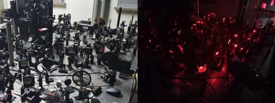
Super-depth 3D holographic microscope. Image Credit: Institute for Basic Science
It is believed that the new microscope can “see-through” the intact skull and is efficient for high-resolution 3D imaging of the neural network inside a living mouse brain without requiring the skull's removal. As a reference, the mouse skull and a human fingernail exhibit identical opacity and thickness.
To scrutinize the interior organs of a living organism with light, it is essential to offer the sample considerable light energy and precisely quantify the signal reflected from the target tissue.
In living tissues, however, multiple scattering effects and extreme aberration are likely to happen as the light falls on the cells, making it tough to achieve sharp images.
In intricate structures like living tissues, the light goes through multiple scattering, causing the photons to alter their direction multiple times at random as they move through the tissue. Much of the image data carried by the light gets damaged as a result.
Despite this issue, with a tiny amount of reflected light it is possible to observe the structures situated comparatively deep inside the tissues by correcting the wavefront distortion of the light reflected from the to-be-observed target.
The above-mentioned multiple scattering results, however, obstruct this process of correction. Hence, to gain a high-resolution deep-tissue image, it is essential to eliminate the multiple-scattered waves and raise the single-scattered waves’ ratio.
In 2019, the IBS scientists made the high-speed time-resolved holographic microscope for the first time. It can remove multiple scattering and, at the same time, quantify the amplitude and light phase. They employed this microscope to witness the neural network of live fish without the need for incisional surgery.
Conversely, when considering a mouse with a relatively thicker skull than a fish, it is impossible to achieve an image of the brain’s neural network without thinning or removing the skull, owing to extreme light distortion and multiple scattering happening while the light passes across the bone structure.
The study group succeeded in quantitatively analyzing the interaction between light and matter, which enabled them to improve their previous microscope even more.
This latest research noted the fruitful development of a super-depth, three-dimensional time-resolved holographic microscope that enables tissue observation to a better depth than ever before.
In particular, the researchers developed an approach to favorably choose single-scattered waves due to the fact that they exhibit identical reflection waveforms even while the light is given from several angles.
This process is performed by a complicated algorithm and a numerical function that examines the eigenmode of a medium (a unique wave that offers light energy into a medium), allowing the result of a resonance mode that increases constructive interference (interference that happens during the overlapping of the same phase waves) between wavefronts of light.
This allowed the new microscope to concentrate more than 80 times of light energy on the neural fibers than before while eliminating unwanted signals selectively. This enabled an increase of several orders of magnitude in the ratio of single-scattered waves versus multiple-scattered waves.
The study group established this new technology by witnessing the mouse brain. The microscope could correct the wavefront distortion even at a previously considered impossible depth with the current technology.
The new microscope successfully achieved a high-resolution image of the neural network of the mouse brain present within the skull. All was attained in the visible wavelength without removing the mouse skull and necessitating a fluorescent label.
“When we first observed the optical resonance of complex media, our work received great attention from academia. From basic principles to practical application of observing the neural network beneath the mouse skull, we have opened a new way for brain neuroimaging convergent technology by combining the efforts of talented people in physics, life, and brain science,” said Professor Moonseok Kim and Dr. Yonghyeon Jo, who have made the foundation of the holographic microscope.
Journal Reference
Jo, Y., and Lee, Y-R. (2022) Through-skull brain imaging in vivo at visible wavelengths via dimensionality reduction adaptive-optical microscopy. Science Advances. doi.org/10.1126/sciadv.abo4366.
Source: https://www.ibs.re.kr/eng.do
https://www.azooptics.com/News.aspx?newsID=27926

![]()
Tel. +82-62-715-4703~4 / Fax. +82-62-715-4707
Copyright(c) 2014 Center for Relativistic Laser Science at IBS. All Rights Reserved.