mainmenu
Interferometric Scattering Microscopy to Characterize Nanometric Objects and Subcellular Structures: Towards Fast 3D Imaging at Nanoscale.
BPS2020
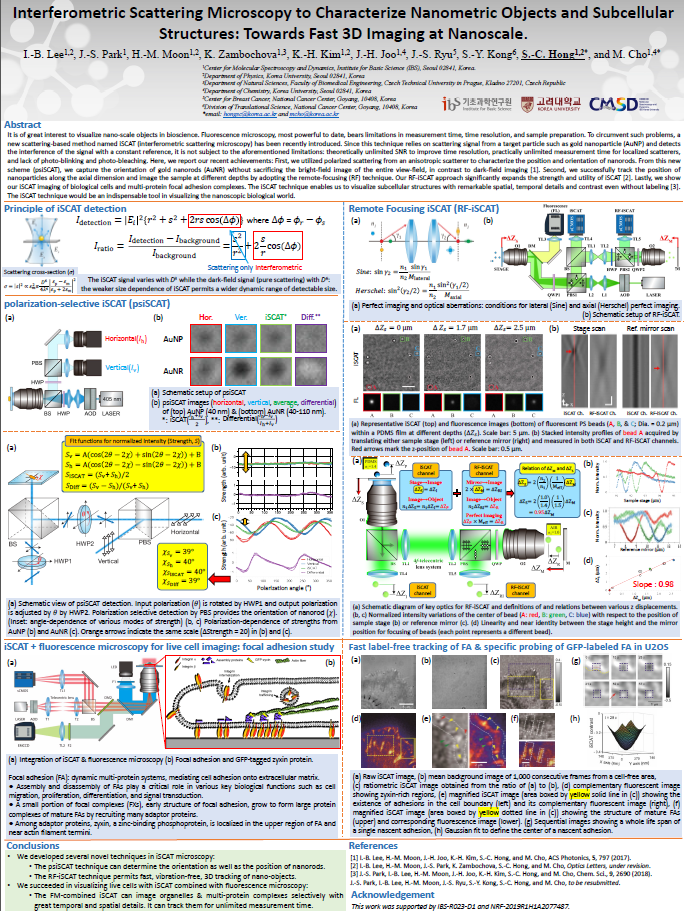
It is of great interest to visualize nano-scale (biological) objects in both nano- and bio-science. Several ingenious methods such as fluorescence-based super-resolution microscopic techniques have been developed to meet the need. These techniques, however, bear evident limitations in measurement time, time resolution, and sample preparation as they mainly rely on fluorescence from fluorescent proteins or artificially introduced fluorescent probes. In order to circumvent such problems, a new scattering-based method named iSCAT (interferometric scattering microscopy) has been recently introduced. Since this technique relies on scattering signal from a target particle and detects the interference of the signal with a constant reference, it is, in principle, not subject to the aforementioned limitations: theoretically unlimited signal-to-noise ratio to improve time resolution, practically unlimited measurement time for stably localized scatterers, and lack of photophysical and photochemical artefacts such as photo-blinking and photo-bleaching.
In this presentation, first, we would like to show our recent application of this technique to image biological cells. Interestingly, the iSCAT technique enables us to visualize subcellular structures with remarkable spatial, temporal resolutions and contrast without any labeling [1].
Second, in order to characterize the position and orientation of nanorods, we utilized scattering of polarized light from anisotropic scatterers, the strength of which is a function of relative orientation of the scatterers to the direction of polarization. From this new scheme named psiSCAT (polarization selective iSCAT), we successfully demonstrate that one can capture the orientational information of anisotropic nanorods without sacrificing the bright-field image of the entire view-field, which is unavailable for dark-field imaging [2].
Third, we are able to track the position of nanoparticles along the axial dimension and to image the sample at different depth with minimal image distortion by adopting the remote-focusing technique. We believe that the adoption of the remote-focusing approach significantly expands the strength and utility of iSCAT [3].
This new scattering-based microscopy technique would be an indispensable tool in visualizing the nanoscopic material world and would shed new light on phenomena occurring in the nano-space.
This work was supported by IBS-R023-D1 and NRF-2019R1H1A2077487.
References
[1] J.-S. Park, I.-B. Lee, H.-M. Moon, J.-H. Joo, K.-H. Kim, S.-C. Hong, and M. Cho, Chem. Sci., 9, 2690 (2018). J.-S. Park, I.-B. Lee, H.-M. Moon, J.-S. Ryu, S.-Y. Kong, S.-C. Hong, and M. Cho, submitted.
[2] I.-B. Lee, H.-M. Moon, J.-H. Joo, K.-H. Kim, S.-C. Hong, and M. Cho, ACS Photonics, 5, 797 (2017).
[3] I.-B. Lee, H.-M. Moon, J.-S. Park, K. Zambochova, S.-C. Hong, and M. Cho, in preparation.

![]()
Tel. +82-62-715-4703~4 / Fax. +82-62-715-4707
Copyright(c) 2014 Center for Relativistic Laser Science at IBS. All Rights Reserved.