mainmenu
Z sectioning of intracellular organelles with remote-focusing-enabled iSCAT microscopy
2019.07 Single-molecule biophysics conference
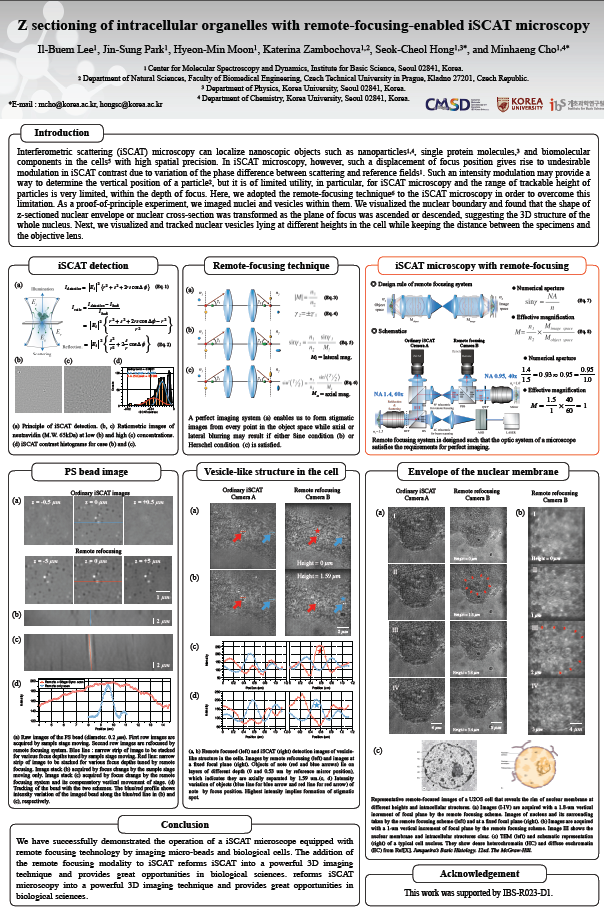
Interferometric scattering (iSCAT) microscopy can localize nanoscopic objects such as nanoparticles1,4, single protein molecules,3 and biomolecular components in the cells5 with high spatial precision. It also enables us to observe the spatial organization of subcellular organelles, dynamical behaviors and co-localization of vesicles, promising great potential in biological research. Three-dimensional image of living cells and organisms can be achieved by optical sectioning at various depths along the optical axis by changing the distance between the specimens and the objective lens. In iSCAT microscopy, however, such a displacement of focus position gives rise to undesirable modulation in iSCAT contrast due to variation of the phase difference between scattering and reference fields1, and thus the relative phase between scattering and reflection fields must remain constant in time and irrespective of the depth of focus. Such an intensity modulation may provide a way to determine the vertical position of a particle2, but it is of limited utility, in particular, for iSCAT microscopy and the range of trackable height of particles is very limited, within the depth of focus.
Here, we adopted the remote-focusing technique6 to the iSCAT microscopy in order to overcome this limitation. The remote-focusing technique is an ideal approach here because the relative phase between the fields remains the same and the plane of focus can be swiftly swept for fast 3D imaging because a light mirror, not heavy and bulky objective, is translated for focus change. As a proof-of-principle experiment, we imaged nuclei and vesicles within them. First we visualized the nuclear boundary and found that the shape of z-sectioned nuclear envelope or nuclear cross-section was transformed as the plane of focus was ascended or descended, suggesting the 3D structure of the whole nucleus. Next, we visualized and tracked nuclear vesicles lying at different heights in the cell while keeping the distance between the specimens and the objective lens.
In summary, we successfully demonstrated that our remote-focusing-equipped iSCAT microscope can track three dimensional objects such as cells along the z-direction over a much larger range of 10 um, which reforms iSCAT microscopy into a powerful 3D imaging technique and provides great opportunities in biological sciences.
[1] K. Lindfors, T. Kalkbrenner, P. Stoller, and V. Sandoghdar. “Detection and Spectroscopy of Gold Nanoparticles Using Supercontinuum White Light Confocal Microscopy.” Phys. Rev. Lett. 93, 297–4 (2004).
[2] Madhavi Krishnan, Nassiredin Mojarad, Phillip Kukura, and Vahid Sandoghdar. Geometry-induced electrostatic trapping of nanometric objects in a fluid. Nature 467, 692–695 (2010).
[3] Marek Piliarik and Vahid Sandoghdar. “Direct optical sensing of single unlabelled proteins and super-resolution imaging of their binding sites.” Nat Commun 5, 4495–8 (2014).
[4] Il-Buem Lee, Hyeon-Min Moon, Jong-Hyeon Joo, Kyoung-Hoon Kim, Seok-Cheol Hong, and Minhang Cho. “Interferometric Scattering Microscopy with Polarization-Selective Dual Detection Scheme: Capturing the Orientational Information of Anisotropic Nanometric Objects.” ACS Photonics 5, 797–804 (2017).
[5] Jin-Sung Park, Il-Buem Lee, Hyeon-Min Moon, Jong-Hyeon Joo, Kyoung-Hoon Kim, Seok-Cheol Hong, and Minhaeng Cho. “Label-free and live cell imaging by interferometric scattering microscopy.” Chem. Sci. 9, 2690–2697 (2018).
[6] Edward J. Botcherby, Rimas Juskaitis, Martin J. Booth, and Tony Wilson. “Aberration-free optical refocusing in high numerical aperture microscopy.” Opt. Lett., OL 32, 2007–2009 (2007).

![]()
Tel. +82-62-715-4703~4 / Fax. +82-62-715-4707
Copyright(c) 2014 Center for Relativistic Laser Science at IBS. All Rights Reserved.