mainmenu
Complementarily balanced real-time two-color SRS
2019 IBS AOI Conference
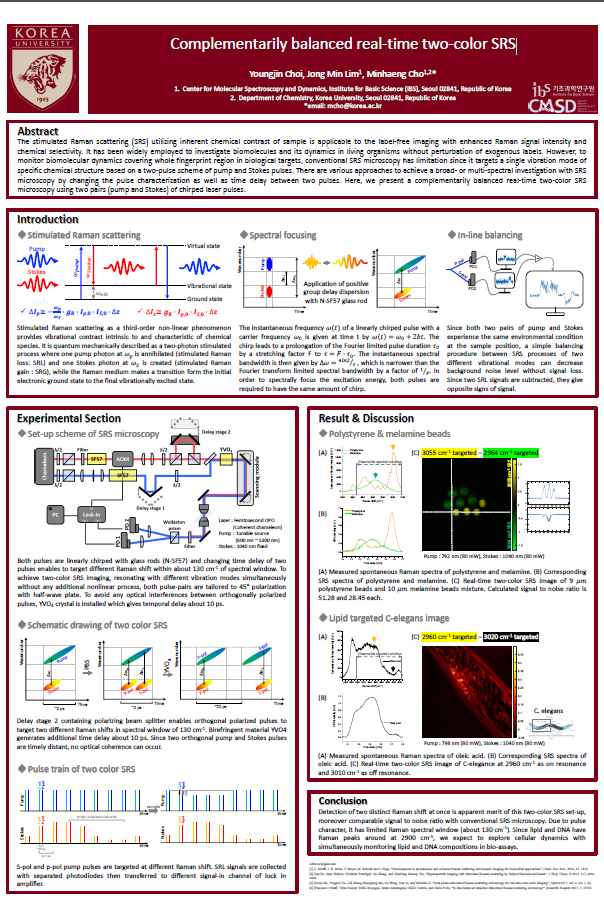
The stimulated Raman scattering (SRS) utilizing inherent chemical contrast of sample is applicable to the label-free imaging with enhanced Raman signal intensity and chemical selectivity. It has been widely employed to investigate biomolecules and its dynamics in living organisms without perturbation of exogenous labels1. However, to monitor biomolecular dynamics covering whole fingerprint region in biological targets, conventional SRS microscopy has limitation since it targets a single vibration mode of specific chemical structure based on a two-pulse scheme of pump and Stokes pulses. There are various approaches to achieve a broad- or multi-spectral investigation with SRS microscopy by changing the pulse characterization as well as time delay between two pulses.
Here, we present a complementarily balanced real-time two-color SRS microscopy using two pairs (pump and Stokes) of chirped laser pulses. Both pulses are linearly chirped with glass rods (N-SF57) and changing time delay of two pulses enables to target different Raman shift within about 130 cm-1 of spectral window2. To achieve two-color SRS imaging, resonating with different vibration modes simultaneously without any additional nonlinear process, both pulse-pairs are tailored to 45° polarization and temporally (~10 ps) divided into orthogonal polarization to each other3. Since both polarization of pump and Stokes experience the same environmental condition at the sample position, a simple balancing procedure between SRS processes of two different vibrational modes can decrease background noise level without signal loss4. A mixture of melamine and polystyrene beads is investigated targeting on the NH and CH stretching mode at 2964 and 3055 cm-1, respectively. Two distinctive Raman images are detected at the same time and works as off-resonance signals to each other. With this technique, we expect to explore cellular dynamics with simultaneously monitoring lipid and DNA compositions in bio-assays.
References
[1] C. Krafft, I. W. Schie, T. Meyer, M. Schmitt and J. Popp, “Developments in spontaneous and coherent Raman scattering microscopic imaging for biomedical appications”, Chem. Soc. Rev., 2016, 45, 1819.
[2] Dan Fu, Gary Holtom, Christian Freudiger, Xu Zhang, and Xiaoliang Sunney Xie, “Hyperspectral imaging with stimulated Raman scattering by chirped femtosecond lasers”, J. Phys. Chem. B 2013, 117, 4634-4640.
[3] Ruoyu He, Yongkui Xu, Lili Zhang, Shenglong Ma, Xu Wang, Dan Ye, and Minbiao Ji, “Dual-phase stimulated Raman scattering microscopy for real-time two-color imaging”, Optica 2017, vol. 4, No. 1, 44.
[4] Francesco Crisafi, Vikas Kumar, Tullio Scopigno, Marco Marangoni, Giulio Cerullo, and Dario Pollo, “In-line balanced detection stimulated Raman scattering microscopy”, Scientific Reoprts 2017, 7, 10745.

![]()
Tel. +82-62-715-4703~4 / Fax. +82-62-715-4707
Copyright(c) 2014 Center for Relativistic Laser Science at IBS. All Rights Reserved.