mainmenu
Single-molecule Localization Expansion Microscopy
2019 IBS AOI Conference
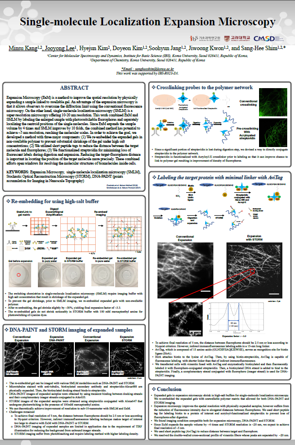
Expansion Microscopy (ExM) is a method to improve the spatial resolution by physically expanding a sample linked to swellable gel. An advantage of the expansion microscopy is that it allows observers to overcome the diffraction limit using the conventional fluorescence microscopy. On the other hand, single-molecule localization microscopy (SMLM) is a super-resolution microscopy offering 10-20 nm resolution. This work combined ExM and SMLM by labeling the enlarged sample with photoswitchable fluorophores and separately localizing the centroid positions of the single molecules. Since ExM expands the sample volume by 4 times and SMLM improves by 10 folds, the combined method has potential to achieve ~5 nm resolution, reaching the molecular scales. In addition to development of imaging method, we develop new labeling method to reduce the distance between the target molecule and fluorophores. Reducing the target-fluorophore distance is important in locating the position of the target molecule more precisely. These combined efforts open windows for resolving the molecular structures of biomolecules inside cells.

![]()
Tel. +82-62-715-4703~4 / Fax. +82-62-715-4707
Copyright(c) 2014 Center for Relativistic Laser Science at IBS. All Rights Reserved.