mainmenu
Mid-IR Photothermal Microscopy
2019 IBS AOI Conference
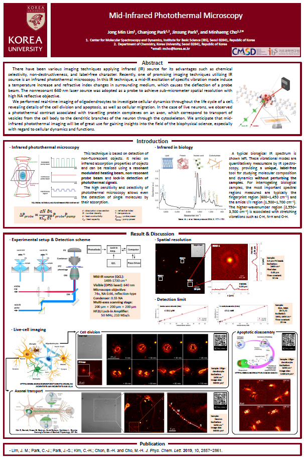
There have been various imaging techniques applying infrared (IR) source for its advantages such as chemical selectivity, non-destructiveness, and label-free character. Recently, one of promising imaging techniques utilizing IR source is an infrared photothermal microscopy. 1,2 In this IR technique, a mid-IR excitation of specific vibration mode induce a temperature increase and refractive index changes in surrounding medium, which causes the deflection of a probe beam. The nonresonant 640 nm laser source was adopted as a probe to achieve sub-micrometer spatial resolution with high NA reflective objective.
We performed real-time imaging of oligodendrocytes to investigate cellular dynamics throughout a life cycle of cell, revealing details of the cell division and apoptosis, as well as cellular migration.3 In the case of live neurons, we observed a photothermal contrast associated with travelling protein complexes on an axon, which correspond to transport vesicles from the cell body to the dendritic branches of the neuron through the cytoskeleton. We anticipate that mid-IR photothermal imaging will be of great use for obtaining insights in the field of the biophysical science, especially regarding cellular dynamics and functions.
References
[1] D. Zhang, C. Li, C. Zhang, M. N. Slipchenko, G. Eakins, J.-X. Cheng, “Depth-resolved mid-infrared photothermal imaging of living cells and organisms with submicrometer spatial resolution.”, Sci. Adv. 2016, 2, e1600521.
[2] Z. Li, K. Aleshire, M. Kuno, G. V. Hartland, “Super-Resolution Far-Field Infrared Imaging by Photothermal Heterodyne Imaging.”, J. Phys. Chem. B 2017, 121, 8838-8846.
[3] J. M. Lim, C.-J. Park, J.-S. Park, C.-H. Kim, B.-H. Chon, and M.-H. Cho, “Cytoplasmic Protein Imaging with Mid-Infrared Photothermal Microscopy: Cellular Dynamics of Live Neurons and Oligodendrocytes”, J. Phys. Chem. Lett. 2019, 10, 2857−2861.

![]()
Tel. +82-62-715-4703~4 / Fax. +82-62-715-4707
Copyright(c) 2014 Center for Relativistic Laser Science at IBS. All Rights Reserved.