mainmenu
Real-time observation of the individual focal adhesion dynamics and cytoskeletons via iSCAT microscopy
2019 IBS AOI Conference
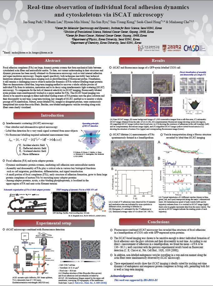
Focal adhesion complexes (FAs) are large, dynamic protein systems that form mechanical links between cytoskeletal actin fibers and extracellular matrix. To date, our current understanding in their structures and dynamic processes has been mostly obtained via fluorescence microscopy such as total internal reflection and super-resolution microscopy. Despite superb specificity, both techniques inevitably bear technical limitations inherent in fluorescence imaging such as photobleaching of fluorescent probes. Nonetheless, it still remains a challenging issue to observe molecular dynamics of FAs without labelling target proteins. Here we demonstrate a label-free, long-term imaging method to uncover a whole cellular process of individual FAs from its initiation, maturation and to its decay using interferometric light scattering (iSCAT) microscopy.1-3 To compensate for the lack of chemical selectivity in iSCAT imaging, fluorescently-labeled zyxin proteins were simultaneously tracked as a major marker for FAs. The iSCAT-based imaging was shown to be sensitive enough to detect individual binding events of FA proteins onto the glass substrate and their disassembly in real time. Long-term tracking, key strength of iSCAT, enabled us to monitor a whole passage of FA metabolism. Mature, zyxin-labeled FAs, imaged as elongated patches, were continuously interpolated into zyxin-free actin fibers. Catastrophic response of fibers and FAs to latrunculin A, actin polymerization inhibitor, vividly imaged in real time, gave insights into the identity of fibers and their coupling to FAs. Besides, non-labeled endogenous vesicles traveling along actin fibers were clearly visualized. These experimental results show that the iSCAT-based method is ideally suited for tracking real-time dynamics of endogenous and exogenous protein complexes in living cells, permitting both fast as well as long-term imaging.
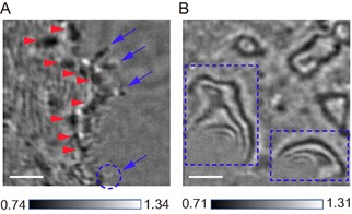
Figure 1. iSCAT image of COS-7 cell boundaries from Ref [4]. (A) Close contact areas of a COS-7 cell spreading onto a glass substrate. (B) Strong fringe patterns formed along the cell boundary. The scale bar is 2 mm.
References
[1] K. Lindfors, T. Kalkbrenner, P. Stoller, and V. Sandoghdar, “Detection and spectroscopy of gold nanoparticles using supercontinuum white light confocal microscopy”, Phys. Rev. Lett. 2004, 93, 037401.
[2] J. O. Arroyo, D. Cole, and P. Kukura, “Interferometric scattering microscopy and its combination with single-molecule fluorescence imaging”, Nat. Protoc. 11, 617-633 (2016).
[3] R. W. Tayler, R. G. Mahmoodabadi, V. Rauschenberger, A. Giessl, A. Schambony, and V. Sandoghdar, “Interferometric scattering microscopy reveals microsecond nanoscopic protein motion on a live cell membrane”, Nat. Photonics 2019, https://doi.org/10.1038/s41566-019-0414-6.
[4] J.-S. Park, I.-B. Lee, H.-M. Moon, J-H. Joo, K.-H. Kim, S.-C. Hong, and M. Cho, “Label-free and live cell imaging by interferometric scattering microscopy”, Chem. Sci. 2018, 9, 2690.

![]()
Tel. +82-62-715-4703~4 / Fax. +82-62-715-4707
Copyright(c) 2014 Center for Relativistic Laser Science at IBS. All Rights Reserved.