mainmenu
Diffusion dynamics of fluorescent nano-diamonds in living cells detected by fluorescence-combined iSCAT system
2019 IBS AOI Conference
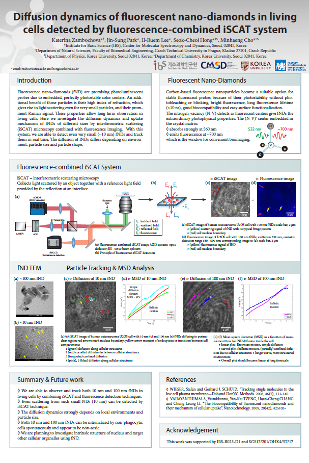
Fluorescent nano-diamonds (fNDs) are promising photoluminescent probes due to embedded, perfectly photostable color centers. These carbon-based fluorescent nanoparticles became a suitable option for stable fluorescent probes in bioimaging applications because of their bright fluorescence, long fluorescence lifetime, no photobleaching, biocompatibility and easy surface functionalization1,2,3. As a downside, the intrinsic number of fluorescent centers in a nanodiamond is relatively low4 and the concentration of the centers decreases nonlinearly with decreasing crystal size5. An additional benefit of those particles is the high index of refraction6, which gives rise to light scattering even for very small particles. Combination of these properties allow long-term observation in living cells.
Here we investigated the diffusion dynamics and uptake mechanisms of fNDs of different sizes in real time by interferometric scattering (iSCAT) microscopy7 combined with fluorescence imaging. While at short-term observation the particles exhibit Brownian motion, the long-term measurement shows directionality in fND diffusion inside the cell. The diffusion of fNDs differs depending on surrounding local environments, particle size and particle shape. The diffusion of 10 nm fNDs resembles that of naturally occurring vesicles in cells. On the other hand, 100 nm fNDs progress faster and move straight towards the cell nucleus. Additionally, we observed spontaneous endocytosis of 10 nm fND without any functional treatment on the surface in real time.
In conclusion, we were able to observe and track both 10 nm and 100 nm fNDs in living cells by combining iSCAT and fluorescence detection techniques, indicating that scattering from such small NDs (10 nm) can be detected by iSCAT technique. The diffusion dynamics strongly depends on local environments and particle size. Moreover, both fNDs can be internalized by cells spontaneously and appear to be non-toxic.
References
[1] VAIJAYANTHIMALA, Vairakkannu, Yan-Kai TZENG, Huan-Cheng CHANG and Chung-Leung LI. “The biocompatibility of fluorescent nanodiamonds and their mechanism of cellular uptake”. Nanotechnology. 2009, 20(42), 425103-
[2] WENG, Mao-Feng, Bo-Jui CHANG, Su-Yu CHIANG, Niann-Shiah WANG and Huan NIU. “Cellular uptake and phototoxicity of surface-modified fluorescent nanodiamonds”. Diam. Relat Mater. 2012, 22, 96-104
[3] FU, Chi-Cheng, Hsu-Yang LEE, Kowa CHEN, et al. “Characterization and application of single fluorescent nanodiamonds as cellular biomarkers”. Proc. Natl. Acad. Sci. USA 2007, 104(3), 727-732
[4] HUI, Yuen Yung, Chia-Liang CHENG and Huan-Cheng CHANG. “Nanodiamonds for optical bioimaging”. J. Phys. D. 2010, 43(37), 374021-
[5] BRADAC, Carlo, Torsten GAEBEL, Nishen NAIDOO, James R. RABEAU and Amanda. S. BARNARD. “Prediction and Measurement of the Size-Dependent Stability of Fluorescence in Diamond over the Entire Nanoscale”. Nano Lett. 2009, 9(10), 3555- 3564
[6] BRADAC, Carlo, Torsten GAEBEL, Nishen NAIDOO, James R. RABEAU and Amanda. S. BARNARD. “Prediction and Measurement of the Size-Dependent Stability of Fluorescence in Diamond over the Entire Nanoscale”. Nano Lett. 2009, 9(10), 3555- 3564
[7] J.-S. Park, I.-B. Lee, H.-M. Moon, J-H. Joo, K.-H. Kim, S.-C. Hong, and M. Cho, “Label-free and live cell imaging by interferometric scattering microscopy”, Chem. Sci. 2018, 9, 2690.

![]()
Tel. +82-62-715-4703~4 / Fax. +82-62-715-4707
Copyright(c) 2014 Center for Relativistic Laser Science at IBS. All Rights Reserved.