mainmenu
Aberration-free and flexible bundle-fiber endomicroscopy
2019 IBS AOI Conference
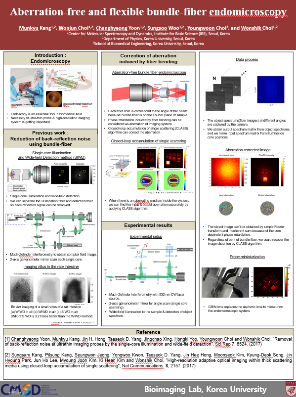
The bundled fiber is commonly used as a probe of endoscopes due to its small dimension, flexibility and relatively large numerical aperture (NA). However, image pixelation and sample-induced aberration degrade spatial resolving power. T-matrix measurement has been proposed to resolve such problems, but it is vulnerable for the fiber bending. Here we apply our own algorithm, Closed-Loop Accumulation of Single Scattering (CLASS)1), to the fiber bundle system for correcting the core-to-core phase retardation induced by fiber bending as well as sample-induced aberration. Back-reflection problem from the ultrathin probe was also solved since we performed the single-core scanning.2) We could demonstrate high-resolution and pixelation-free images. We are in the process of miniaturizing the endomicroscope by attaching a GRIN lens to the bundle-fiber to acquire in-vivo biosample images.
Figures
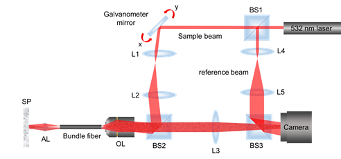
Figure 1. Experimental setup. BS1~BS3: Beam splitters, L1~L5: Lenses, OL: 10x, 0.25 NA objective lens, AL: Effective focal length 1.45 mm aspheric lens. Obtaining complex field images from Mach-Zehnder interferometry system. Scanning each single core of bundle-fiber by swinging the galvanometer mirror. Fiber is placed at the Fourier plane of the sample to measure the aberration of core dependent aberration.
References
[1] Kang. S, et al., “High-resolution adaptive optical imaging within thick scattering media using closed-loop accumulation of single scattering”, Nat. Communications. 8. 2157. (2017).
[2] Yoon. C, et al., “Removal of back-reflection noise at ultrathin imaging probes by the single-core illumination and wide-field detection”, Scientific Reports. 7, 6524. (2017).

![]()
Tel. +82-62-715-4703~4 / Fax. +82-62-715-4707
Copyright(c) 2014 Center for Relativistic Laser Science at IBS. All Rights Reserved.