mainmenu
Selective Activation DNA-PAINT Imaging with Reductive Caging
2018 KCS 122nd
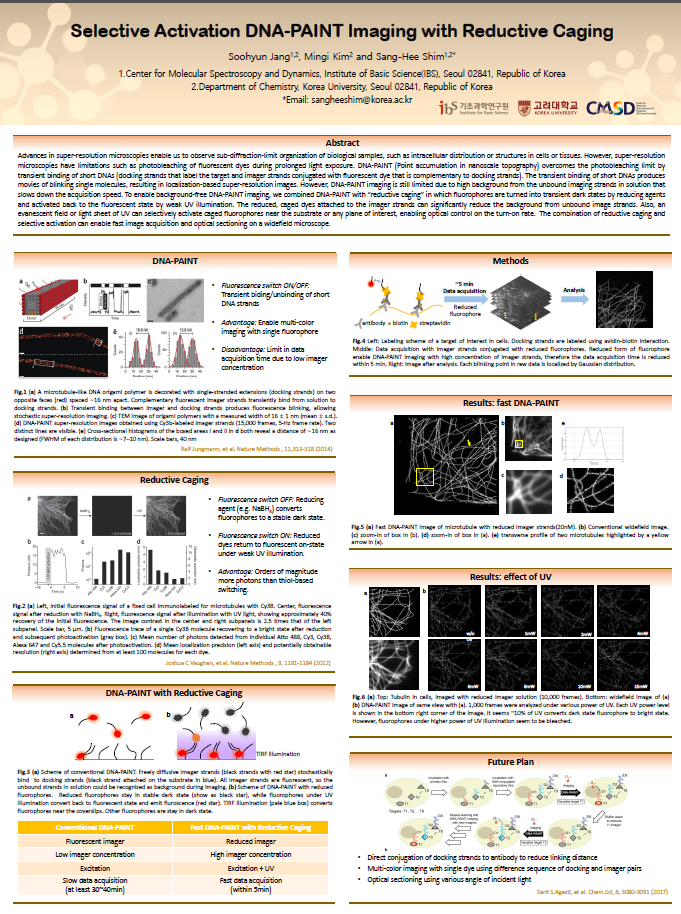
Advances in super-resolution microscopies enable us to observe sub-diffraction-limit organization of biological samples, such as intracellular distribution or structures in cells or tissues. However, super-resolution microscopies have limitations such as photobleaching of fluorescent dyes during prolonged light exposure. DNA-PAINT (Point accumulation in nanoscale topography) overcomes the photobleaching limit by transient binding of short DNAs (docking strands that label the target and imager strands conjugated with fluorescent dye that is complementary to docking strands). The transient binding of short DNAs produces movies of blinking single molecules, resulting in localization-based super-resolution images. However, DNA-PAINT imaging is still limited due to high background from the unbound imaging strands in solution that slows down acquisition speed. To enable fast DNA-PAINT imaging, we combined DNA-PAINT with “reductive caging” in which fluorophores are turned into transient dark states by reducing agents and activated back to the fluorescent state by weak UV illumination. The reduced, caged dyes attached to the imager strands can significantly reduce the background from unbound image strands. Also, an evanescent field of UV can selectively activate caged fluorophores near the substrate, enabling optical control on the turn-on rate. The combination of reductive caging and selective activation can effectively reduce the image acquisition time in a controllable manner.
References
[1] Vaughan, J., Jia, S. and Zhuang, X. (2012). Ultrabright photoactivatable fluorophores created by reductive caging. Nature Methods, 9(12), pp.1181-1184.

![]()
Tel. +82-62-715-4703~4 / Fax. +82-62-715-4707
Copyright(c) 2014 Center for Relativistic Laser Science at IBS. All Rights Reserved.