mainmenu
High-resolution and label-free neuroimaging of a living zebrafish by simultaneous angular scanning microscopy
ABC 2018
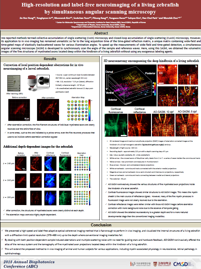
We reported methods termed collective accumulation of single scattering (CASS) microscopy and closed-loop accumulation of single scattering (CLASS) microscopy. However, its application to in vivo imaging has remained unrealistic so far to the long acquisition time of the time-gated reflection matrix, a unique matrix containing wide-field and time-gated maps of elastically backscattered waves for various illumination angles. To speed up the measurements of wide-field and time-gated detection, a simultaneous angular scanning microscope (SASM) is developed to synchronously scan the angle of the sample and reference waves. Here, using this SASM, we obtained the volumetric images of the fine structures of myelinated axons located deep within the hindbrain of a living zebrafish without using any exogenous labeling agents.
REFERENCE
Kang, S. et al. (2015) Nat. Photonics 9, 253–258.
Kang, S. et al. (2017) Nat. Communications 8, 2157.

![]()
Tel. +82-62-715-4703~4 / Fax. +82-62-715-4707
Copyright(c) 2014 Center for Relativistic Laser Science at IBS. All Rights Reserved.