mainmenu
Label-free and high-resolution adaptive optical microscopy for deep-tissue imaging of a living zebrafish
2018 GRC
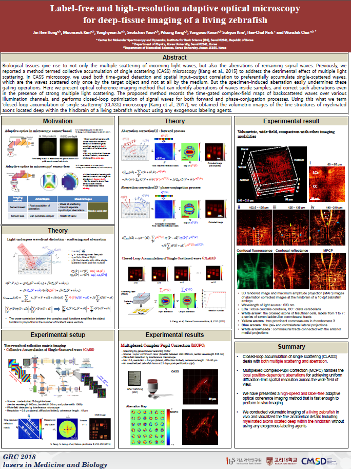
Biological tissues give rise to not only the multiple scattering of incoming light waves, but also the aberrations of remaining signal waves. Previously, we reported a method termed collective accumulation of single scattering (CASS) microscopy [Kang et al., 2015] to address the detrimental effect of multiple light scattering. In CASS microscopy, we used both time-gated detection and spatial input–output correlation to preferentially accumulate single-scattered waves, which are the waves scattered only once by the target object and not at all by the medium. But the specimen-induced aberration easily undermines these gating operations. Here we present optical coherence imaging method that can identify aberrations of waves inside samples, and correct such aberrations even in the presence of strong multiple light scattering. The proposed method records the time-gated complex-field maps of backscattered waves over various illumination channels, and performs closed-loop optimization of signal waves for both forward and phase-conjugation processes. Using this what we term ‘closed-loop accumulation of single scattering’ (CLASS) microscopy [Kang et al., 2017], we obtained the volumetric images of the fine structures of myelinated axons located deep within the hindbrain of a living zebrafish without using any exogenous labeling agents.
REFERENCE
Kang, S. et al. (2015) Nat. Photonics 9, 253–258.
Kang, S. et al. (2017) Nat. Communications 8, 2157.

![]()
Tel. +82-62-715-4703~4 / Fax. +82-62-715-4707
Copyright(c) 2014 Center for Relativistic Laser Science at IBS. All Rights Reserved.