mainmenu
Label-free Microscopy Using Vibrational Resonance by Stimulated Raman Scattering
2018 121st KCS General Meeting
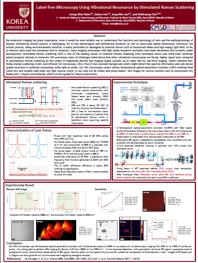
Abstract: Bio-molecular imaging has great importance, since it would be most reliable way to understand the functions and physiology of cells and the pathophysiology of disease. Monitoring cellular process is challenging, for its fast physiological and biochemical dynamics as well as nano-scale spatial dimensions. Moreover, the cellular process, being environmentally sensitive, is easily perturbed (or damaged) by external stimuli such as fluorescent labels and high energy light field. So far, to observe nano-scale bio-complexes and its dynamics, many imaging techniques with high spatio-temporal resolution have been developed and currently under development.
Stimulated Raman scattering (SRS) is one of the leading classes of coherent Raman scattering (CRS) technique which uses third-order nonlinear optical property intrinsic to molecule.
SRS overcomes many of challenges listed above that other vibrational microscopies are facing. Higher signal level compared to spontaneous Raman scattering by few orders of magnitude permits fast imaging speed, possibly up to video rate for real-time imaging. Unlike coherent anti-Stokes Raman scattering (CARS) and infrared (IR) microscope, SRS is free of non-resonant background which might distort the spectral information and sub-micron spatial resolution is achieved conducting visible light as probe. Our SRS microscope system utilizes femtosecond optical-parametric oscillator (OPO) emitting fixed (1040 nm) and tunable (680-1040 nm) light sources which, in our case will be Stokes and pump beams. Test images for various samples such as polystyrene (PS) beads and C. Elegans are achieved, which will be a guide for future experiments.

![]()
Tel. +82-62-715-4703~4 / Fax. +82-62-715-4707
Copyright(c) 2014 Center for Relativistic Laser Science at IBS. All Rights Reserved.