mainmenu
Raman and Fluorescence Imaging with Polymer Dot
2018 121st KCS General Meeting
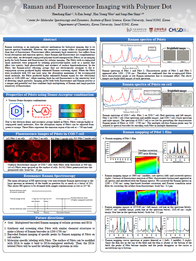
Raman scattering is an emerging contrast mechanism for biological imaging due to its narrow spectral bandwidth. However, the sensitivity is many orders of magnitude lower than that of fluorescence. Fluorescence offers extremely high sensitivity, but suffers from broad absorption and emission spectra. Since Raman and fluorescence are complementary to each other, we developed conjugated polymer-based nanoparticles (Pdots) as an imaging probe for both Raman and fluorescence for cellular imaging. The Pdots with π-conjugated small molecules were prepared by utilizing poly(styrene-maleic acid) as a matrix that offers low toxicity, high photostability and easiness of surface functionalization. In the Pdots, the Raman-active vibrating groups that are electronically resonant to the π-conjugation system produced highly enhanced Raman scattering signal. When the Pdots were irradiated with 532 nm laser near the absorption maximum of the π-conjugated small molecule, the Pdot produced highly enhanced Raman signal for the vibrational modes at 1200-1800 cm-1, while emitting high far-red fluorescence to the sensitivity level of detecting single particles. Imaging probe with both fluorescence and Raman-activity is a unique and powerful tool that combines high multiplexing of Raman and single-particle sensitivity of fluorescence.

![]()
Tel. +82-62-715-4703~4 / Fax. +82-62-715-4707
Copyright(c) 2014 Center for Relativistic Laser Science at IBS. All Rights Reserved.