mainmenu
High-resolution adaptive optical imaging within thick scattering media using closed-loop accumulation of single scattering
2018 FOM
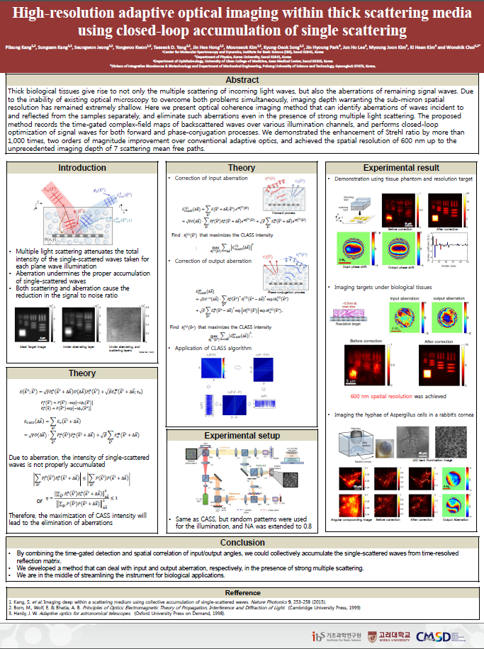
1. Abstract
When considering a target spatial resolution close to the ultimate diffraction limit, the attenuation of SNR by multiple light scattering is not the only problem. In fact, the specimen-induced aberration is an equally important issue to address [1]. The signal waves that preserve original incidence momenta are not only attenuated in their intensity by the multiple light scattering, but their phases are also retarded due to the heterogeneity of the medium. These phase retardations vary depending on the propagation angle [2].
In this presentation, we present a label-free and high-resolution imaging method that can identify sample-induced aberrations in illumination and imaging paths separately even in the presence of strong multiple light scattering. We used a time-gated optical coherence imaging to record the amplitude and phase maps of backscattered waves from the specimens for various illumination angles. In the image reconstruction process, we introduced separate angle-dependent phase factors for the incident and reflected waves, and identified phase corrections that preferentially accumulate single-scattered waves over multiple-scattered ones for the forward and phase-conjugation processes. By applying these angle-dependent phase corrections to the initial data, we could significantly reduce the effect of image distortion. Using this what we term ‘closed-loop accumulation of single scattering’ (CLASS) microscopy, we achieved a spatial resolution of 600 nm up to the imaging depth of 7 scattering mean free paths, an unprecedented imaging depth at such a high resolving power. To demonstrate the applicability of CLASS microscopy to biological tissues, we conducted imaging of a rabbit’s cornea infected by the aspergillus fumigatus, a type of fungi, and successfully visualized individual fungal filaments therein.
2. References
[1]Hell, S., Reiner, G., Cremer, C. & Stelzer, E. H. Aberrations in confocal fluorescence microscopy induced by mismatches in refractive index. Journal of microscopy 169, 391-405 (1993).
[2]Wyant, J. C. & Creath, K. Basic wavefront aberration theory for optical metrology. Applied optics and optical engineering 11, 2 (1992).
[3]Kang, S., Jeong, S., Choi, W., Ko, H., Yang, T. D., Joo, J., Lee, J., Lim, Y., Park, Q. & Choi, W. Imaging deep within a scattering medium using collective accumulation of single-scattered waves Nature Photonics 9, 253-258 (2015)

![]()
Tel. +82-62-715-4703~4 / Fax. +82-62-715-4707
Copyright(c) 2014 Center for Relativistic Laser Science at IBS. All Rights Reserved.