mainmenu
Live-cell Imaging with Interferometric Scattering Microscopy (iSCAT)
124th Summer Symposium of KCS-Physical Chemistry Division
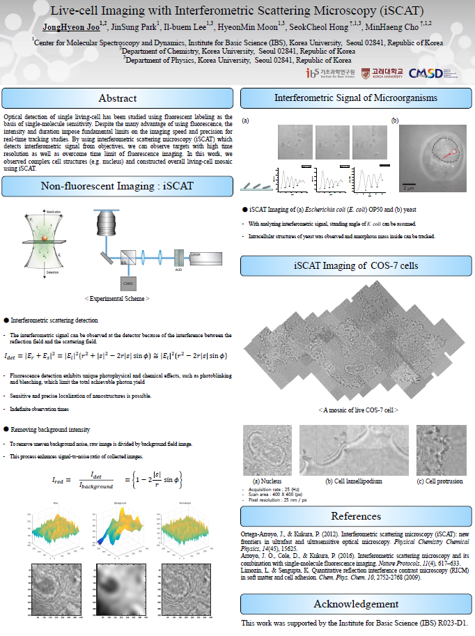
Optical detection of single living-cell has been studied using fluorescent labeling as the basis of single-molecule sensitivity. Despite the many advantage of using fluorescence, the intensity and duration impose fundamental limits on the imaging speed and precision for real-time tracking studies. By using interferometric scattering microscopy (iSCAT) which detects interferometric signal from objectives, we can observe targets with high time resolution as well as overcome time limit of fluorescence imaging. In this work, we observed complex cell structures (e.g. nucleus) and constructed overall living-cell mosaic using iSCAT.
References
[1] Ortega-Arroyo, J., & Kukura, P. (2012). Interferometric scattering microscopy (iSCAT): new frontiers in ultrafast and ultrasensitive optical microscopy. Physical Chemistry Chemical Physics, 14(45), 15625.
[2] Arroyo, J. O., Cole, D., & Kukura, P. (2016). Interferometric scattering microscopy and its combination with single-molecule fluorescence imaging. Nature Protocols, 11(4), 617–633.

![]()
Tel. +82-62-715-4703~4 / Fax. +82-62-715-4707
Copyright(c) 2014 Center for Relativistic Laser Science at IBS. All Rights Reserved.