mainmenu
Highly Bright Expansion Microscopy
121st Summer Symposium of KCS-Physical Chemistry Division (2016)
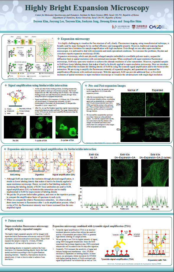
Expansion microscopy (ExM), which uses physically enlarged samples embedded in a swellable polymer matrix, surpass the diffraction limit in spatial resolution with conventional microscopes. When combined with super-resolution fluorescence microscopy, ExM may open new windows to achieve the ultimate resolution of a few nanometers. However, expanded samples in ExM have lower labeling density that makes it hard to be directly applied to super-resolution microscopy. Here we introduce a labeling method that increases the labeling density of ExM by using the tyramide signal amplification and/or the biotin/avidin interaction. We amplified the fluorescence intensity of expanded samples, and quantitatively examined the amplification efficiencies with confocal microscopy. With this approach, ExM can provide additional four- or five-fold increment of spatial resolution in super-resolution microscopy to visualize the ultrastructures with single-digit resolution.
References
[1] F. Chen, P. W. Tillberg and E. S. Boyden, Science, 347, 543 (2015).
[2] T. J. Chozinski, A. R. Halpern, H. Okawa, H. J. Kim, G. J. Tremel, R. O. L. Wong and J. C. Vaughan, Nature Methods,13, 485 (2016).

![]()
Tel. +82-62-715-4703~4 / Fax. +82-62-715-4707
Copyright(c) 2014 Center for Relativistic Laser Science at IBS. All Rights Reserved.